Nano-Tracking Plaques
Researchers from Adlershof and the Charité, Europe’s largest university clinic, are working on advancing the use of element microscopes
Modern medical imaging aims at looking into the human body and assessing the condition of the circulatory system and organs as accurately as possible. It deals with potentially life-saving questions. Is a heart attack or stroke imminent due to arteriosclerosis, i.e. thickening blood vessels through build-up? Researchers from the BAM Federal Institute for Materials Research and Testing and Charité have developed a special microscope to look into these questions.
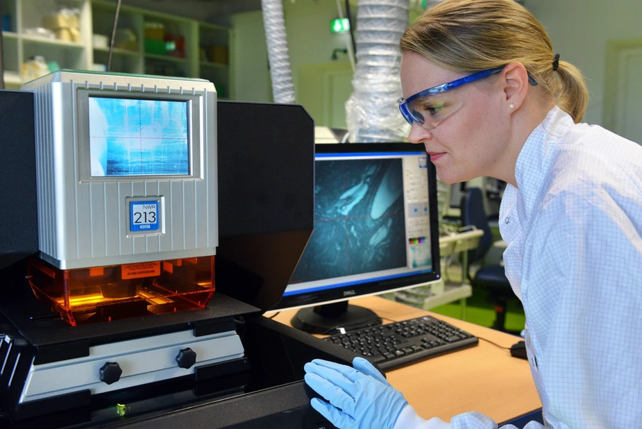
Using intricate procedures, Charité professor Eyk Schellenberger, head of the working group Molecular Imaging, and his staff track tiny objects on the nanoscale: exogenous markers or endogenous atoms and molecules. They developed a special element microscope in cooperation with a team led by Norbert Jakubowski, head of the inorganic trace analysis division of the BAM Federal Institute for Materials Research and Testing in Adlershof.
“It helps us to measure the nanoparticle distribution in clinical tissue samples,” says Jakubowski. They can provide crucial information on pathogenic processes. However, it is hard to produce high-res images of these changes. This issue is resolved using markers, which are attached to cells to send out luminous or magnetic signals. The latter are emitted by very small iron oxide particles, VSOPs for short, which are injected into blood vessels and traced using magnetic resonance imaging (MRI). “It takes about an hour until these reach the arteriosclerotic plaques,” says Schellenberger. After attaining a PhD at Halle University in 1999, the professor specialised in molecular imaging research at Harvard Medical School and has now been teaching and researching at the university clinic in Berlin since 2004.
To safely identify the VSOPs, the researchers at Charité mix them with Europium, a rare earth metal, which is rare in the human body. “A Europium content of 0.3 percent does not affect the properties of the VSOP,” he says. How does one accurately analyse the sample taken from a lab mouse and visualise the europium-based VSOPs?
This is where the element microscope comes into play, which Schellenberger first became aware of at a conference. This instrument serves to trace and analyse individual elements in biological tissue sections. Looking for Berlin-based specialists in this field, he met Jakubowski and his colleague Larissa Müller, a chemist. The resulted in the joint project between Charité and BAM, which is being funded by the German Research Foundation. Today, PhD student Lena Ascher is working on optimising the procedure, which involves vaporising tissue samples and breaking them down into their individual elements in the plasma of a special mass spectrometer. It enables researchers to measure the quantity of europium and iron in the plaques located the mouse’s aorta with very high accuracy.
Currently, one measurement of the element microscope yields data on 30 parameters, including markers and antibodies. It lets us look ever more closely into the human body.
By Paul Janositz for Adlershof Journal
