Battery research with the HZB X-ray microscope
Experiments at BESSY II have clarified chemical changes in lithium-ion batteries
New cathode materials are being developed to further increase the capacity of lithium batteries. Multilayer lithium-rich transition metal oxides (LRTMOs) offer particularly high energy density. However, their capacity decreases with each charging cycle due to structural and chemical changes. Using X-ray methods at BESSY II, teams from several Chinese research institutions have now investigated these changes for the first time with highest precision: at the unique X-ray microscope, they were able to observe morphological and structural developments on the nanometre scale and also clarify chemical changes.
Lithium-ion batteries are set to become even more powerful with new materials for the cathodes. For example, layered lithium-rich transition metal (LRTMO) cathodes could further increase the charge capacity and be used in high-performance lithium batteries. However, so far it has been observed that these cathode materials ‘age’ rapidly: the cathode material degrades as a result to the back-and-forth migration of lithium ions during charging and discharging. Until now it was unclear what specific changes these would involve.
Teams from Chinese research institutions have therefore applied for beam time at the world's only transmission X-ray microscope (TXM) at an undulator beamline at the BESSY II storage ring to investigate their samples using 3D tomography and nanospectroscopy. The HZB-TXM measurements were performed by Dr. Peter Guttmann, HZB, back in 2019, before the coronavirus pandemic. The X-ray microscopic analysis was then supplemented by further spectroscopic and microscopic examinations. After careful evaluation of the extensive data, the results are now available: they provide detailed information on changes in the morphology and structure of the material, but also on chemical processes during discharge.
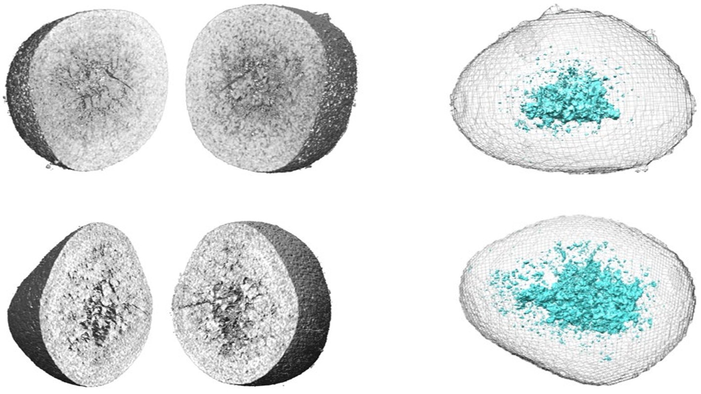
‘Soft X-ray transmission microscopy allows us to visualise chemical states in LRTMO particles in three dimensions with high spatial resolution and to gain insights into chemical reactions during the electrochemical cycle,’ explains Dr Stephan Werner, who is responsible for the scientific supervision and further development of the instrument.
The results provide insights into local lattice distortions associated with phase transitions and nanopore formation. The oxidation states of individual elements could also be determined locally. The speed of the charging processes plays an important role here: slow charging favours phase transitions and oxygen loss, while fast charging leads to lattice distortions and inhomogeneous lithium diffusion.
‘Here at the TXM, we have a unique capability: we can offer energy-resolved transmission X-ray tomography,’ says Werner. ’This gives us a 3D image with structural information at every element-specific energy level – energy is the fourth dimension here.’
The results from this study provide valuable information for the development of high-performance cathodes that remain stable over the long term and are resistant to cycling. ‘The TXM is excellently suited to provide new insights into morphological and chemical changes in battery materials in the future through in-operando studies – that is, during charging and discharging,’ says Prof. Gerd Schneider, who developed the TXM.
Publication:
Nature Nanotechnology (2024): Revealing the degradation pathways of layered Li-rich oxide cathodes
Zhimeng Liu, Yuqiang Zeng, Junyang Tan, Hailong Wang, Yudong Zhu, Xin Geng, Peter Guttmann, Xu Hou, Yang Yang, Yunkai Xu, Peter Cloetens, Dong Zhou, Yinping Wei, Jun Lu, Jie Li, Bilu Liu, Martin Winter, Robert Kostecki, Yuanjing Lin & Xin He
DOI: 10.1038/s41565-024-01773-4
Contact:
Helmholtz-Zentrum Berlin für Materialien und Energie
Department X-Ray Microscopy
Dr. Stephan Werner
+49 30 8062-13181
Email
Prof. Dr. Gerd Schneider
+49 30 8062-13131
Email
Press Officer:
Dr. Antonia Rötger
+49 30 8062-43733
Email
Press release HZB, 18 November 2024